Thursday, October 11, 2012

Block 2 Case 20 Answers

A 70 year old man presents to the doctor for a routine physical. When you listen to his heart you hear the following:
What kind of murmur does he have? Aortic stenosis

What is the most likely cause of his murmur? calcified deposits due to "wear and tear"

It this a typical finding in a 70 year old man? No, typically the murmur would be heard in a man over 80. The fact that you are hearing the murmur now suggests that he has a bicuspid aortic valve. 

Name 4 symptoms associated with this murmur

  1. fatigue
  2. dyspnea on exertion
  3. angina
  4. syncope
  5. sudden cardiac death
If he spiked a fever after having dental work what would you be concerned of? Strep viridans subacute endocarditis

If he had an aortic valve replacement what type of infective endocarditis would he be more at risk of contracting? Staph. Epidermidis is found in patients who have had a valve replacement. He could also still get Acute bacterial endocarditis (Staph. Aureus) or Subacute bacterial endocarditis (Strep Viridans).

http://emedicine.medscape.com/article/2018199-overview#aw2aab6c10

Wednesday, October 10, 2012

Block 2 - case 20


A 70 year old man presents to the doctor for a routine physical. When you listen to his heart you hear the following:
What kind of murmur does he have?

What is the most likely cause of his murmur? 

It this a typical finding in a 70 year old man?

Name 4 symptoms associated with this murmur

If he spiked a fever after having dental work what would you be concerned of?

If he had an aortic valve replacement what type of infective endocarditis would he be more at risk of contracting? 


Tuesday, October 9, 2012

Block 2 Case 19 Answers


A 12 year old female comes to your office with her mother because of a slightly raised painless rash which developed yesterday morning.  She notes swollen joints, palpitations, and jerky uncontrolled movements.  You discover that she is visiting from Argentina and had a sore throat and fever 3 weeks prior.  She has a temperature of 103.1 in the office today.


Below are tissue sections from heart biopsy.


What key features are present in the biopsy specimens?
1    1)Aschoff Body – lymphocytes, occasionally plasma cells, and activated macrophages
2)     Anitschkow cells (pathognomonic for RF)  - activated histiocytes which have abundance cytoplasm and a central, slender, wavy, ribbon of chromatin.  May become multinucleated.  Characteristic “caterpillar chromatin.”
3)     Fibrinoid necrosis, often perivascular
4)     Verrucaw along the lines of valve closure overlying areas of fibrinoid necrosis within the cusps or along the chordae tendinae


What is the diagnosis?
Acute Rheumatic Fever

What is the pathogenesis?
Systemic illness following Group A beta hemolytic strep pharyngitis.  Due to cross-reactivity of antibodies to strep antigens with host cardiac antigens resulting in cardiac damage.  ARF results from immune response to group A strep.   

What are the Jones criteria for RF? What do you need for diagnosis?

Major Criteria:
1.      Migratory polyarthralgias
2.      Pancarditis
3.      Sydenham’s Chorea
4.      Erythema Marginatum
5.      Subcutaneous Nodules
Minor:
            Fever, Arthralgia, elevated ESR,  CRP, PR, WBC

Dx: 2 major or 1 major + 2 minor + evidence of group a strep infection

What organism would grow following cardiac culture?
            None.  Culture negative. 


The patient returned after several more episodes of rheumatic heart disease and died from complications from heart failur.  Autopsy specimens are shown below:


What key findings are present in the autopsy specimens?
  •   Leaflet thickening
  • 2 Thickening and fusion of the chordae tendinae


What valves are typically affected in rheumatic heart disease?
            Mitral> Mitral + Aortic > Aortic Alone


Block 2 Case 19


A 12 year old female comes to your office with her mother because of a slightly raised painless rash which developed yesterday morning.  She notes swollen joints, palpitations, and jerky uncontrolled movements.  You discover that she is visiting from Argentina and had a sore throat and fever 3 weeks prior.  She has a temperature of 103.1 in the office today.


Below are tissue sections from heart biopsy.


What key features are present in the biopsy specimens?
1   

What is the diagnosis?


What is the pathogenesis?


What are the Jones criteria for RF? What do you need for diagnosis?

Major Criteria:

Minor:
      

What organism would grow following cardiac culture?
    

The patient returned after several more episodes of rheumatic heart disease and died from complications from heart failure.  Autopsy specimens are shown below:


What key findings are present in the autopsy specimens?

What valves are typically affected in rheumatic heart disease?


Block 2 Case 17 Answers


Below is an image from a biopsy of a 17 year old athlete who developed chest pain and dyspnea during a tennis match and passed out. He was rushed to the hospital but was dead on arrival.
http://www.sciencedirect.com/science/article/pii/S1756231709001960

What are the key histologic findings?
   -Interstitial fibrosis


There is a genetic component to this disease  What is the mutation? Is it autosomal dominant or recessive?


Hypertrophic cardiomyopthy is genetic in many cases: autosomal dominant with mutations in genes of sarcomere proteins
   -35% beta-myosin heavy chain
   -15% myosin binding protein C

What was the cause of his symptoms?
  • Due to a septal obstruction and poor compliance there was reduced filling during diastole which lead to a lower cardiac output that was unable to provide sufficient oxygen to vital organs.  Insufficient oxygen to the brain lead to syncope. Inadequate oxygenation of his heart caused his syncope.
  • Dyspnea was due to the stifened left  ventricle which impaired blood flow resulting in pressures that backed up fluid into the lungs.


Block 2 Case 17


Above is an image from a biopsy of a 17 year old athlete who developed chest pain and dyspnea during a tennis match and passed out. He was rushed to the hospital but was dead on arrival.
http://www.sciencedirect.com/science/article/pii/S1756231709001960

What are the key histologic findings?

There is a genetic component to this disease. What is the mutation?

 Is it autosomal dominant or recessive?

What was the cause of his symptoms?

Block 2 Case 16 Answers


1. In Los Angeles a 38 yo HIV positive man who recently traveled to Peru and originally complaining of fatigue is hospitalized with palpitations and dyspnea. He has no significant medical history and does not take any medications. He has a 20 pack year smoking history and reports occasional alcohol use, though his wife mentions that it is daily. He denies illicit drug use.

Labs:
Hemoglobin 14 g/dL
MCV 101
AST 55 U/L
ALT 45 U/L
TSH 4.5uU/mL

On Physical exam, his temp is 98.5F, BP is 120/80mmHg, and heart rate is 115/min. Jugular venous pressure is normal. The lungs are clear. Cardiac examination shows an irregularly irregular rhythm. There is trace edema at both ankles. 

Chest radiograph was as follows:

b. Based on his history and clinical presentation, what will you find on the echocardiogram that the Cardiologist ordered on your patient?

Dilated Cardiomyopathy
(For this patient,you found dilated ventricles with normal wall thickness and severely decreased systolic function--left ventricular ejection fraction, 15%)
c. What other pertinent positives in his history leads you to and exacerbates the diagnosis above? What in his labs support your etiology?
Alcoholic History

d.  Why was TSH ordered? What does this patient's TSH mean?

Severe hypothyroidism can cause myxedema heart

e. What other drugs could have cause this patient's symptoms?

Cocaine, Amiodarone, Daunorubicin, Doxorubicin


Ultimately, the patient stopped drinking alcohol to slow down the progression of his illness. The doctor, convinced that there was another related, but undiscovered etiology, sought out a cardiac biopsy ruling almost everything out.
f. What new additional diagnosis do you now have?

Chagas Disease


Block 2 Case 16

1. In Los Angeles a 38 yo HIV positive man who recently traveled to Peru and originally complaining of fatigue is hospitalized with palpitations and dyspnea. He has no significant medical history and does not take any medications. He has a 20 pack year smoking history and reports occasional alcohol use, though his wife mentions that it is daily. He denies illicit drug use.

Labs:
Hemoglobin 14 g/dL
MCV 101
AST 55 U/L
ALT 45 U/L
TSH 4.5uU/mL

On Physical exam, his temp is 98.5F, BP is 120/80mmHg, and heart rate is 115/min. Jugular venous pressure is normal. The lungs are clear. Cardiac examination shows an irregularly irregular rhythm. There is trace edema at both ankles. 

Chest radiograph was as follows:

b. Based on his history and clinical presentation, what will you find on the echocardiogram that the Cardiologist ordered on your patient?

c. What other pertinent positives in his history leads you to and exacerbates the diagnosis above? What in his labs support your etiology?
d.  Why was TSH ordered? What does this patient's TSH mean?

e. What other drugs could have cause this patient's symptoms?

Ultimately, the patient stopped drinking alcohol to slow down the progression of his illness. The doctor, convinced that there was another related, but undiscovered etiology, sought out a cardiac biopsy ruling almost everything out.
f. What new additional diagnosis do you now have?


Block 2 Case 15 Answers


Block 2 Case 15
1. A father and his 15 year old daughter present to you with a chief complaint of primary amenorrhea. In addition, the young prepubescent woman also complains of changes in her toes she has never brought up before.

Upon inspection:


Upon seeing her toes, your clinical suspicion leads you to order a number of tests, including a lateral magnetic resonance angiography of the the thorax.

MRA Result:


a. What is your suspected etiology of the clubbing and cyanosis complaint by your 15 year old patient?

Coarctation of the aorta (preductal)

b. Which genetic abnormality is associated with the above etiology?

60% of cases-Nondisjunction 45, X
40% of cases- Mosaicism 45X, XX Karyotype

c. Of the clinical symptoms listed below, what five are associated with this genetic abnormality.

-Short stature
-Decreased estradiol and progesterone
-Streak gonads
-Increased FSH and LH
-Cystic Hydroma

Block 2 Case 15

1. A father and his 15 year old daughter present to you with a chief complaint of primary amenorrhea. In addition, the young prepubescent woman also complains of changes in her toes she has never brought up before.

Upon inspection:

Upon seeing her toes, your clinical suspicion leads you to order a number of tests, including a lateral magnetic resonance angiography of the the thorax. 

MRA Result:

a. What is your suspected etiology of the clubbing and cyanosis complaint by your 15 year old patient?

b. Which genetic abnormality is associated with the above etiology?

c. Of the clinical symptoms listed below, what five are associated with this genetic abnormality. 

-Tracheoesophageal fistula
-Endocardial cushion defect 
-Short stature
-Lactose intolerance 
-Decreased estradiol and progesterone
-Increased incidence in younger mothers
-History of duodenal atresia at birth
-Heterochromia 
-Panhypopituitarism
-Streak gonads
-Renal failure 
- Increased FSH and LH
-Increased incidence of cystic fibrosis
-Cystic Hydroma


Block 2 Case 14 Answers


In clinic, you meet a new patient who just transferred clinics due to insurance. You are struck by how tan his skin seems to be after noticing the difference between the younger picture of himself in his chart as a younger man. To your surprise, he mentions he has not been able to see the Phlebotomist as often as he usually does.


a. Based on the following liver pathology sample stained in Prussian Blue, what genetic condition does this patient likely have? Name the clinical finding you noticed on his skin. Hereditary Hemochromatosis/Bronze Diabetes


b. Looking through his chart, you find there is also a significant piece of family history. You notice previous work ups and note the findings of a cardiac biopsy report, show below. Name the disease process and one major cardiac clinical finding. Familial Amyloidosis + Arrythmia/Heart Failure

This section of myocardium demonstrates amorphous deposits of pale pink material between myocardial fibers. This is characteristic for amyloid. 

c. You also see this second pathology report. What type of staining did they do and summarize the findings.

A Congo red stain has been performed on the myocardium in a case of amyloidosis. The amyloid stains orange-red, but with polarized light, the amyloid has an "apple-green" birefringence as seen here.

d. What disease process do these two conditions have in common? Restrictive Cardiomyopathy

e. What the pathophysiology behind this disease process? Name one of two.

-Decreased ventricular compliance
-Diastolic dysfunction type of LHF

f. Name two other etiologies of this disease process

Most Common:
-Amyloidosis, 
-Radiation
-Myocardial fibrosis after open heart surgery
Infiltrative
-Hemochromatosis
-Pompe's glycogenosis
Endocardial Fibroelastosis in a child
-thick fibroelastic tissue in the endocardium
Sarcoidosis
Systemic Sclerosis


Block 2 Case 14

In clinic, you meet a new patient who just transferred clinics due to insurance. You are struck by how tan his skin seems to be after noticing the difference between the younger picture of himself in his chart as a younger man. To your surprise, he mentions he has not been able to see the Phlebotomist as often as he usually does.


a. Based on the following liver pathology sample stained in Prussian Blue, what genetic condition does this patient likely have? Name the clinical finding you noticed on his skin. 


b. Looking through his chart, you find there is also a significant piece of family history. You notice previous work ups and note the findings of a cardiac biopsy report, show below. Name the disease process and one major cardiac clinical finding. 


c. You also see this second pathology report. What type of staining did they do and summarize the findings.

d. What disease process do these two conditions have in common? 

e. What the pathophysiology behind this disease process? Name one of two.

f. Name two other etiologies of this disease process


Block 2 Case 13 Answers


1. A concerned couple comes into pediatric clinic with their new adopted baby who noticed that recently when the baby starts to cry, she becomes more fussy. They started to notice this around the baby's 3 month mark. Now, approaching 4 months old, the baby seems to be more irritable, moving more, and has now started to have episodes where her lips, fingers, and toes turn blue.

A chest radiograph shows the following:

a. Based on your clinical suspicion, why do you think the infant has become increasingly fussy when crying? Name the congenital heart defect and the pathophysiology behind the infants reaction.

Tetralogy of Fallot
-Caused by sudden increase in hypoxemia and cyanosis
-Movement increases systemic vascular resistance, causing temporary reversal of the shunt.

b. Using the two images, identify the two congenital heart defects which would increase oxygenation in this infant. Of the two congenital heart defect presented, which can be manipulated at birth through pharmacological intervention?

PDA- through prostaglandins

Congenital Heart Defect 1 -ASD
Congenital Heart Defect 2 -PDA

c. After identifying the four anomalies that make up the Tetralogy of Fallot, name the one whose severity correlates with presence or absence of cyanosis.

-Ventricular Septal Defect
-Infundibular or valvular pulmonary stenosis
-Right ventricular hypertrophy
-Destrorotated aorta with right-sided aortic arch

Block 2 Case 13

1. A concerned couple comes into pediatric clinic with their new adopted baby who noticed that recently when the baby starts to cry, she becomes more fussy. They started to notice this around the baby's 3 month mark. Now, approaching 4 months old, the baby seems to be more irritable, moving more, and has now started to have episodes where her lips, fingers, and toes turn blue.

A chest radiograph shows the following:

a. Based on your clinical suspicion, why do you think the infant has become increasingly fussy when crying? Name the congenital heart defect and the pathophysiology behind the infants reaction.


b. Using the two images, identify the two congenital heart defects which would increase oxygenation in this infant. Of the two congenital heart defect presented, which can be manipulated at birth through pharmacological intervention?

Congenital Heart Defect 1 
Congenital Heart Defect 2 

c. After identifying the four anomalies that make up the Tetralogy of Fallot, name the one whose severity correlates with presence or absence of cyanosis.

Block 2 Case 12 Answers


 1. An infant of uncertain dates is born via emergent cesarean section after the mother was critically injured in a motor vehicle accident. Birth weight was 1075 g. The infant has poor respiratory effort and you begin bag-mask ventilation but find it extremely difficult to cause chest wall movement.

A chest radiograph reveals diffuse whiteout "ground glass appearance" of both lungs.

The following picture demonstrates the underlying pathology.


a. What is the diagnosis?

Respiratory Distress of the Newborn

b. Less than how many weeks is the gestational age of this infant?

Less than 32 or 34 weeks

c. Using the following Electron Mircrograph of a type II pneumocyte name the major pathophysiological reason behind this event, name what is stored inside the object identified by the arrow, and what gestational week it is synthesized..


d. Name three etiologies of this condition.

Prematurity, Maternal Diabetes, and Cesarean Section

e. The treatment for this is CPAP with endotracheal tube with O2 and surfactant. Name 3 of the 5 complications this syndrome can cause:

Superoxide free radical damage from O2 therapy(may result in blindness and permanent damage to small airways)
Intraventricular hemorrhage
Patent ductus arteriosus
Necrotizing enterocolitis
Hypoglycemia in newborn (Excess insulin decreases serum glucose, producing seizures and damage to neurons)


Block 2 Case 12


 1. An infant of uncertain dates is born via emergent cesarean section after the mother was critically injured in a motor vehicle accident. Birth weight was 1075 g. The infant has poor respiratory effort and you begin bag-mask ventilation but find it extremely difficult to cause chest wall movement.

A chest radiograph reveals diffuse whiteout "ground glass appearance" of both lungs.

The following picture demonstrates the underlying pathology.


a. What is the diagnosis?


b. Less than how many weeks is the gestational age of this infant?


c. Using the following Electron Mircrograph of a type II pneumocyte name the major pathophysiological reason behind this event, name what is stored inside the object identified by the arrow, and what gestational week it is synthesized..


d. Name three etiologies of this condition.


e. The treatment for this is CPAP with endotracheal tube with O2 and surfactant. Name 3 of the 5 complications this syndrome can cause:


Block 2 Case 11 Answers


1. "A 28 year female comes to the emergency room complaining of 6 days of fevers with shaking chills. Over the past 2 days, she has also developed a productive cough with greenish sputum, which occasionally is blood streaked. She reports no dyspnea, but sometimes experiences chest pain on deep inspiration.

She denies headache, abdominal pain, urinary symptoms, vomiting, or diarrhea. She smokes cigarettes and marijuana regularly, drinks beers daily, but denis IV drug use.

On exam, her temperature is 102.5 F, heart rate is 109 bpm, bp 128/76 mmHg, and resp rate at 23 breaths per minute. She is alert and talkative. She has no oral lesions, and funduscopic exam reveals no abnormalities. She is tachycardic, but regular with a harsh holosystolic murmur at the left lower sternal border that increases with inspiration. Chest examination revels inspiratory rales bilaterally.


a. What is your clinical suspicion at this point using your clinical gestalt?

Suspected Endocarditis (S. aureus)

Labs:
WBC 17,500/mm3 (Diff: 84% PMN's, 7%bands, 9% lymphs)
Hemoglobin 14 g/dL
Hematocrit 42%
Platelet 189,000/mm3
Liver function and urinalysis are normal

b. What general process are you concerned about after seeing the lab results? Use the following picture. Describe what you can appreciate. 


Infection; 

Microscopically, the valve in infective endocarditis demonstrates friable vegetations of fibrin and platelets (pink) mixed with inflammatory cells and bacterial colonies (blue). The friability explains how portions of the vegetation can break off and embolize.

c. Identify all 4 of the following clinical presentations. Identify the ONE of the four that matches the history of this patient.

Clinical Presentation 1- ROTH SPOTS
-Funduscopic examination


Clinical Presentation 2- TRACK MARKS
-On both of her forearms, you notice this...though you don't notice erythema, warmth, or tenderness.


Clinical Presentation 3-Janeway Lesions
-Non-tender, small erythematous or hemorrhagic macular or nodular lesions on the palms or soles only a few millimeters in diameter

Clinical Presentation 4-OSLERS NODES
-Painful, red, raised lesions found on the hands and feet. 


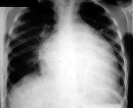
You decide to get a chest radiograph which shows the following:
The radiologist describes them as multiple peripheral, ill-defined nodules, with some cavitation.

d. What is your diagnosis and next step?

Septic Pulmonary Emboli
Obtain serial blood cultures and institute broad spectrum antibiotics. Most likely S. aureus...so treat with Vancomycin.